How AI-Powered Diagnostic Tools Improve Accuracy in Medical Imaging and Pathology
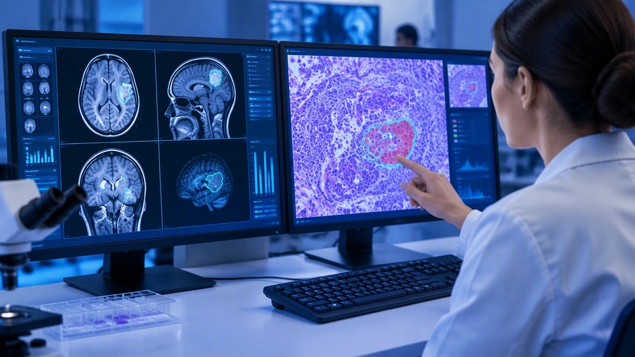
Artificial intelligence is changing how clinicians interpret medical images, but its most useful role is not to replace them. Modern diagnostic systems analyze pixels, tissue patterns, and subtle changes that can be difficult for the human eye to quantify consistently. When carefully developed and validated, these tools can support more accurate readings, faster triage, and earlier detection of disease.
Understanding how this technology works starts with a simple idea: machine learning learns patterns from examples. Instead of following fixed rules written by programmers, a model studies many labeled images and adjusts itself until it can make reliable predictions. This approach is especially valuable in medicine, where complex visual information often contains clues that do not fit neatly into simple thresholds.
How Machine Learning Learns to Read Medical Images
A machine learning model is a mathematical system with many adjustable parameters. During training, it receives an image and produces an output, such as a score indicating the likelihood of a finding. The model’s prediction is compared with the correct answer, and the difference is used to update its parameters. Repeating this process across thousands of examples gradually improves performance.
In deep learning, the model uses multiple layers of connected processing units. Early layers may learn to recognize edges, textures, and contrasts. Deeper layers combine those features into more meaningful patterns, such as a mass, a lesion, or abnormal tissue architecture. The model does not understand medicine in the human sense. It learns statistical relationships that help it classify images or highlight regions that deserve attention.
Medical images come in many forms. Radiology produces X-rays, computed tomography scans, magnetic resonance images, and ultrasound frames. Pathology creates high-resolution images of tissue samples. Ophthalmology captures retinal photographs, while dermatology may use close-up skin images. Each type of data requires its own model design, training set, and evaluation process.
Radiology: Finding Subtle Signs Earlier
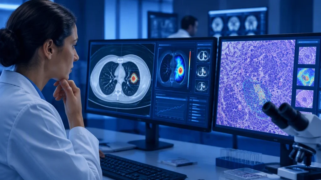
Radiology has become one of the clearest examples of ai in healthcare improving diagnosis accuracy with deep learning. In mammography, for example, software can flag suspicious regions that may indicate early breast cancer. In chest imaging, models can help detect nodules, infections, or signs of lung disease that might be overlooked during a busy shift.
The benefit is not simply speed. A clinician may review hundreds of images in a day, and attention can vary with fatigue, workload, and image quality. An AI system can provide a consistent second review, drawing attention to areas that warrant closer inspection. This kind of support can reduce missed findings while helping radiologists prioritize urgent cases.
Accuracy also depends on context. A model may perform differently across hospitals, scanner brands, patient populations, or image protocols. A system trained mostly on one type of equipment may struggle with another. For this reason, developers test models on independent data and monitor performance after deployment. A useful tool must remain reliable in real clinical conditions, not only in a controlled research setting.
Pathology: Analyzing Tissue at Scale
Pathology relies on detailed examination of tissue samples, often at very high magnification. Digital pathology converts glass slides into whole-slide images that can be reviewed on a screen. Deep learning models can examine these images for patterns associated with cancer, inflammation, or other abnormalities.
One important advantage is quantification. A model can measure the size of a tumor, estimate the proportion of affected tissue, or identify features that are difficult to compare consistently by eye. These measurements may help pathologists describe disease more precisely and support decisions about treatment.
AI can also assist with workflow. In a laboratory handling large volumes of slides, a model can pre-screen samples and identify regions of interest. The pathologist remains responsible for the final interpretation, but the software can reduce repetitive searching and help focus attention where it is most needed.
Early Disease Detection and Screening
Early detection is one of the most promising areas for diagnostic AI. Many diseases produce small changes before symptoms become obvious. In retinal imaging, for instance, models can detect signs of diabetic eye disease from photographs of the back of the eye. In dermatology, image analysis may help evaluate skin lesions. In cardiology, algorithms can assist in identifying abnormalities in heart rhythm or cardiac imaging.
Screening programs create a particular opportunity for improvement because they involve large numbers of people and repeated examinations. An AI system can help standardize the review process and identify individuals who need further testing. However, screening also carries risks. If a model produces too many false alarms, healthy people may undergo unnecessary procedures. If it misses cases, patients may receive a false sense of reassurance.
For this reason, performance is described with more than one number. Sensitivity measures how often the system finds a true abnormality. Specificity measures how often it correctly identifies normal cases. Positive and negative predictive values also depend on how common the disease is in the tested population. A model with impressive accuracy in a laboratory may behave differently in a community clinic where the disease is rare.
What Makes an AI Diagnostic Tool Trustworthy
A trustworthy system begins with high-quality data. Images must be accurately labeled by qualified professionals, and the dataset should represent the patients and settings where the tool will be used. If training data are narrow or biased, the model may fail for certain groups.
Validation is equally important. Developers compare model predictions with expert interpretations and, when available, with clinical outcomes. They examine performance across age groups, sexes, ethnicities, disease stages, and equipment types. Transparent reporting helps clinicians understand what a system can and cannot do.
Human oversight remains essential. AI outputs are best treated as decision support rather than final diagnoses. Clinicians must consider symptoms, medical history, laboratory results, and the patient’s preferences. The software can organize evidence and highlight patterns, but responsibility for care stays with the medical team.
The Path Toward Better Diagnosis
As datasets grow and models become more efficient, diagnostic AI is likely to expand into more specialties and clinical workflows. The greatest gains will come from tools that are not only accurate but also well integrated into everyday practice. Clear interfaces, reliable calibration, and continuous monitoring can help prevent automation bias, where users accept machine output without sufficient scrutiny.
The goal is a balanced partnership. Artificial intelligence can process large amounts of visual information quickly and consistently, while clinicians bring judgment, experience, and an understanding of the whole patient. Together, they can reduce diagnostic errors, detect disease earlier, and make the path from first symptom to appropriate treatment more dependable.